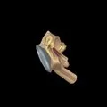
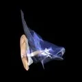
• Origin of the tensor tympani on the temporal
  • Insertion of the tensor tympani on the malleus
  • Origin of the tensor tympani on the skull
  • Origin of the tensor tympani on the auditory ossicles
  • View All
  • 3-D
  • Video
  • Photo
  • Art
  • Histology
  • Cross Section
  • Models
3D - Auditory ossicles with muscle attachments
3D - Malleus with muscle attachments
3D - Muscles of the middle ear (tympanic cavity) inside tympanic cavity
3D - Muscles of the middle ear in glass temporal bone
The right tympanic cavity opened from above. (Enlarged five times.)
The outer wall of the right tympanic cavity as seen from the outer side. (Enlarged five times.)
Illustration - Internal perspective of lateral wall of tympanic cavity
Illustration - Internal perspective of medial wall of tympanic cavity
Dense regular collagenous connective tissue - cross section
Dense regular collagenous connective tissue - longitudinal section
Striated skeletal muscle - longitudinal section
Striated skeletal muscle - cross section
The second branch of the trigeminal nerve, the sphenopalatine ganglion, the intracranial portion of the facial nerve, and the tympanic nerve.
The outer wall of the right tympanic cavity as seen from the inner side. (Enlarged five times.)
The inner wall of the right tympanic cavity seen from the outer side. (Enlarged five times.)
A frontal section of the right external auditory meatus, of the tympanic membrane, and of the tympanic cavity. (Enlarged four times.)
A frontal section of the external auditory meatus, of the tympanic cavity, and the labyrinth. (Enlarged two times.)