• Manubrium of the malleus
  • Head of malleus
  • Neck of the malleus
  • Lateral process of the malleus
  • Anterior process
  • View All
  • 3-D
  • Video
  • Photo
  • Art
  • Histology
  • Cross Section
  • Models
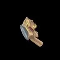
3D - Malleus
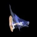
3D - Muscles of the middle ear (tympanic cavity) inside tympanic cavity
3D - Muscles of the middle ear in glass temporal bone
The right malleus from the outer side. (Enlarged seven times.)
The outer wall of the right tympanic cavity as seen from the outer side. (Enlarged five times.)
The outer wall of the right tympanic cavity as seen from the inner side. (Enlarged five times.)
The outer wall of the right tympanic cavity seen from the inner side. (Enlarged five times.)
A frontal section of the right external auditory meatus, of the tympanic membrane, and of the tympanic cavity. (Enlarged four times.)
The right tympanic membrane as seen from the inner side.
Illustration - Frontal section of the sternum and costal cartilages
Illustration - Sternoclavicular and upper costal joints
The right malleus from in front. (Enlarged seven times.)
The right malleus from behind. (Enlarged seven times.)
The right auditory ossicles of a child. (Enlarged seven times.)
Illustration - Sternum
Illustration - Median section of the trunk skeleton
Illustration - Internal perspective of lateral wall of tympanic cavity
Photo - Ear ossicles - size compared to a dime
Photo - Fetal Skull
Photo - Sagittal section through tympanic cavity
Photo - Lateral aspect of malleus
Decalcified bone
Ground bone - Compact bone
Intramembranous bone
The intracranial portion of the facial nerve.
The right tympanic cavity opened from above. (Enlarged five times.)
A frontal section of the external auditory meatus, of the tympanic cavity, and the labyrinth. (Enlarged two times.)